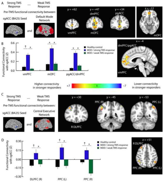
Fig. 4. Baseline subgenual cingulate connectivity predicts treatment response.
A. To test the hypothesis that individual differences in default mode and central executive network connectivity may influence patients’ response to TMS, we compared the pre-treatment functional connectivity maps for patients who subsequently showed a stronger to treatment versus those who showed a weaker response to treatment, based on a median split of patients’ percent change in HAM-D scores. Prior to treatment, patients who subsequently showed larger clinical benefits had higher subgenual cingulate connectivity with multiple nodes of the DMN, including the ventromedial (vmPFC) and dorsomedial prefrontal cortex (dmPFC), pregenual anterior cingulate (pgACC), and medial orbitofrontal cortex (mOFC).
B. Quantification of data extracted from the coordinates of the peak t statistic from each of the areas labeled in panel 4A.
C. Patients who showed a stronger response to TMS also exhibited higher functional connectivity between sgACC, right DLPFC, and bilateral posterior parietal (PPC) areas of the CEN.
D. Quantification of data extracted from the coordinates of the peak t statistic from each of the areas labeled in panel 4C. For coordinates and statistics, see Supplemental Table S7. Quantification of the data depicted in panel 4c. Error bars = S.E.M. * = p < 0.05, corrected for multiple comparisons. † = p < 0.01, uncorrected, but not significant after correcting for multiple comparisons.

