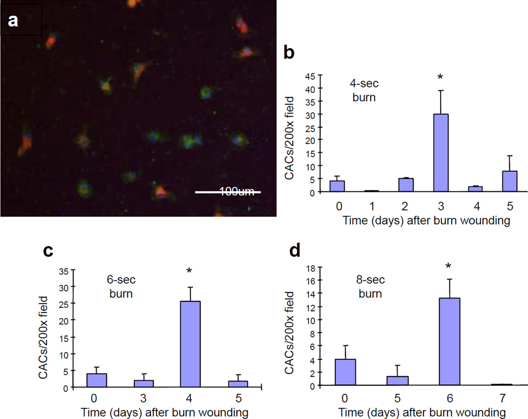
Figure 2. Analysis of circulating angiogenic cells (CACs).
Analysis of CACs in the peripheral blood of mice subjected to burns of increasing duration. Mouse peripheral blood mononuclear cells cultured in the presence of endothelial growth factors were stained with FITC-lectin (green) and DiI-acetylated LDL (red). (a) Mice were subjected to 4-second (b), 6-second (c), and 8-second (d) burns and peripheral blood was analyzed for the presence of CACs on the indicated day after burn wounding (day 0, non-burned controls). *P<0.05 compared to day 0 (Student’s t test).

