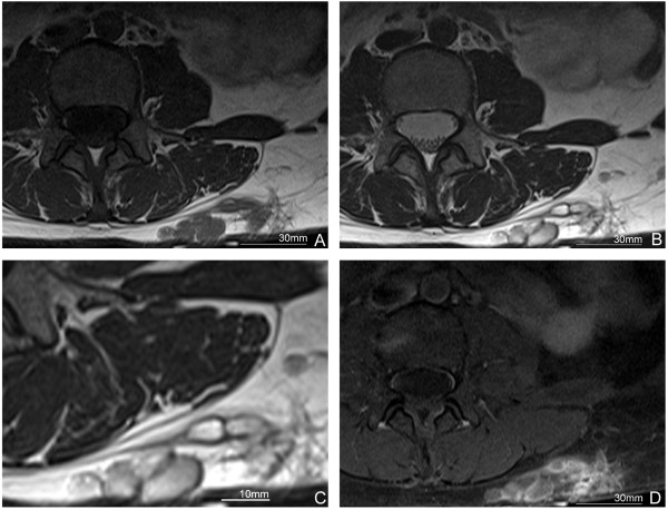
Figure 1.
Magnetic resonance imaging (MRI) findings. A. Polycystic mass with inhomogeneous low intensity on T1-weighted image (T1WI). B. The mass with inhomogeneous high intensity on T2-weighted image (T2WI). C. The magnified image of the nodular lesion in B. D. Rim enhancement is seen but the inside of the mass shows no increment of density after contrast enhancement.

