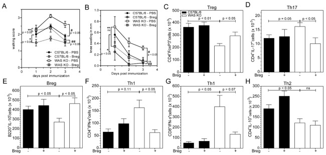
Figure 5.

Transfer of T2 Breg cells ameliorates the development of arthritis in WAS KO mice. (A, B) Severity of experimental arthritis was assessed after Breg-cell transfer as (A) clinical walking score and (B) knee swelling. Data are shown as mean ± SEM from one experiment with control (PBS injected) n = 5, T2 Breg n = 4, #C57BL/6 PBS versus WAS KO PBS, ##C57BL/6 Breg versus WAS KO Breg, *C57BL/6 PBS versus C57BL/6 Breg, **WAS KO PBS versus WAS KO Breg (one-way ANOVA of AUC, p < 0.05). (C–H) The indicated cell populations in the draining LNs of mBSA-injected knees were analyzed by marker staining and flow cytometry. (C) Treg cells, (D) Th17 cells, (E) Breg cells, (F) IFN-γ-expressing CD4+ T cells and (G) CD8+ T cells and (H) IL-10-producing CD4+ T cells were quantified and shown as mean ± SEM of n = 5 (PBS transfer), and n = 4 (T2 Breg-cell transfer) from a single experiment performed. Statistical significance determined by Student's t-test.
