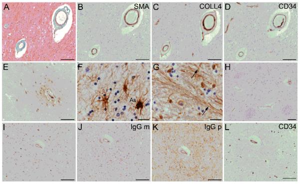
Figure 1.

Histopathological evidence of small vessel disease (SVD) and plasma extravasation in human brain tissue of older people. (A-D) Vascular changes representative of SVD (arteriolosclerosis) in 2 small arteries within subcortical white matter. Masson trichrome stain shows concentric, fibrohyaline thickening due to collagen and other connective tissue (green) in the medial layer, with loss of nuclei (nuclei stained black) (A). The smooth muscle marker α-actin (SMA) confirms partial loss of myocytes from the medial layer. Labeling for collagen-4 (COLL4) shows concentric, “doughnut” labeling, characteristic of small vessel disease (C). CD34 immunolabeling confirms intact endothelium (D). (E) Extravascular fibrinogen is seen around blood vessels in parenchymal cells and as perivascular “collars”. (F, G) Cellular fibrinogen is seen in cells with astrocytic morphology (As) (F), and within neural fibers (arrows, G). (H) Some sections lack extravascular fibrinogen. (I-K) Neighboring sections are immunolabeled for the plasma markers, i.e. fibrinogen (I) or immunoglobulin G monoclonal antibody (IgG m, J) or immunoglobulin G polyclonal antibody (IgG p, K). A neighboring section labeled with anti-CD34 demonstrates endothelial labeling and supports specificity of the labeling with plasma markers. In B-L, the chromogen is 3,3′ diaminobenzidine (brown); the nuclear chromatin counterstain hematoxylin (blue). Panels A-D, F-G, I-L are from subcortical white matter; E and H are from anterior putaminal grey matter. Scale bars: F, G, 10 μm; all other panels, 100 μm.
