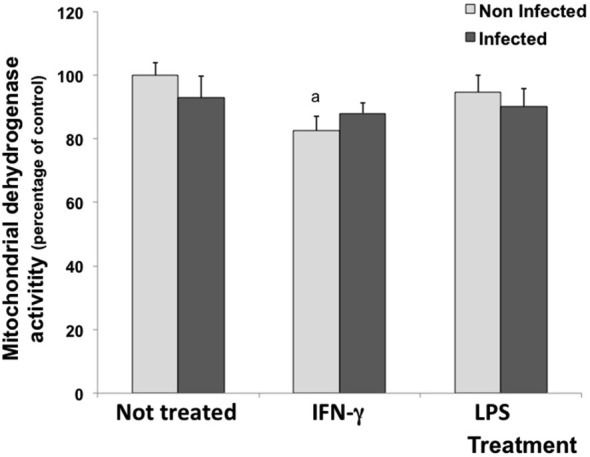
Figure 1.

Percentage of cell viability measured by the MTT assay in rat neuron/glial cell co-cultures treated with 300 IU/mL of IFN-γ and 1.0 mg/mL of LPS and infected with Neospora caninum tachyzoites (ratio cell:parasite 1:1). The results are expressed as the percentage of cell viability observed in different treatment conditions and the respective standard deviation compared with untreated/uninfected control cultures (considered as 100%) 72 h post infection. The results are expressed as the mean of the percentage and the respective standard of eight samples, in three independent experiments. “a” represents a significant statistical difference when compared to untreated/uninfected control cultures; (Two-way ANOVA/Tukey’s Multiple Comparison Test—p < 0.05).
