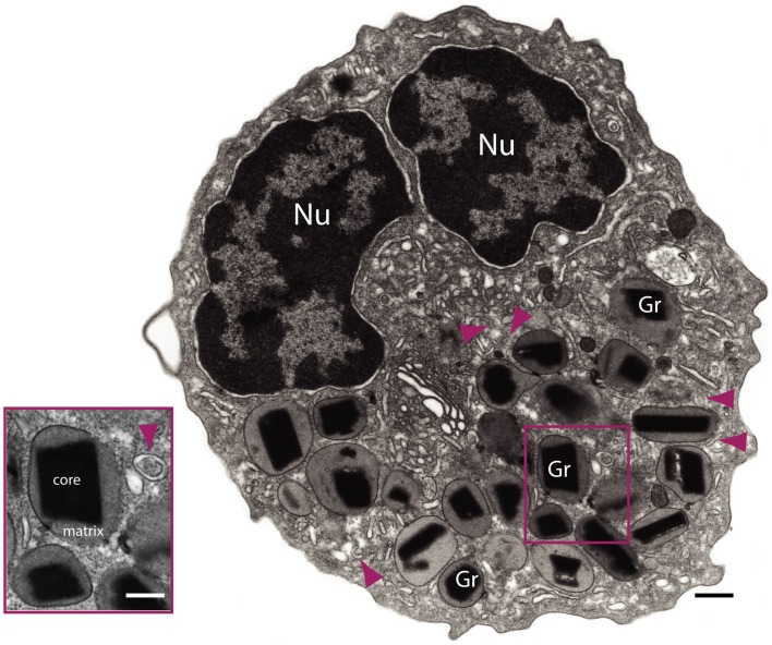
Figure 1.
Transmission electron microscopy of a human eosinophil. This cell is characterized by a major population of specific granules (Gr) with a unique morphology – an internal often electron-dense crystalline core and an outer electron-lucent matrix surrounded by a delimiting trilaminar membrane. Note the typical bilobed nucleus (Nu) and large tubular carriers (arrowheads). The inset shows secretory granules and a tubular vesicle at higher magnification. Bars: 500 nm; 300 nm (inset).

