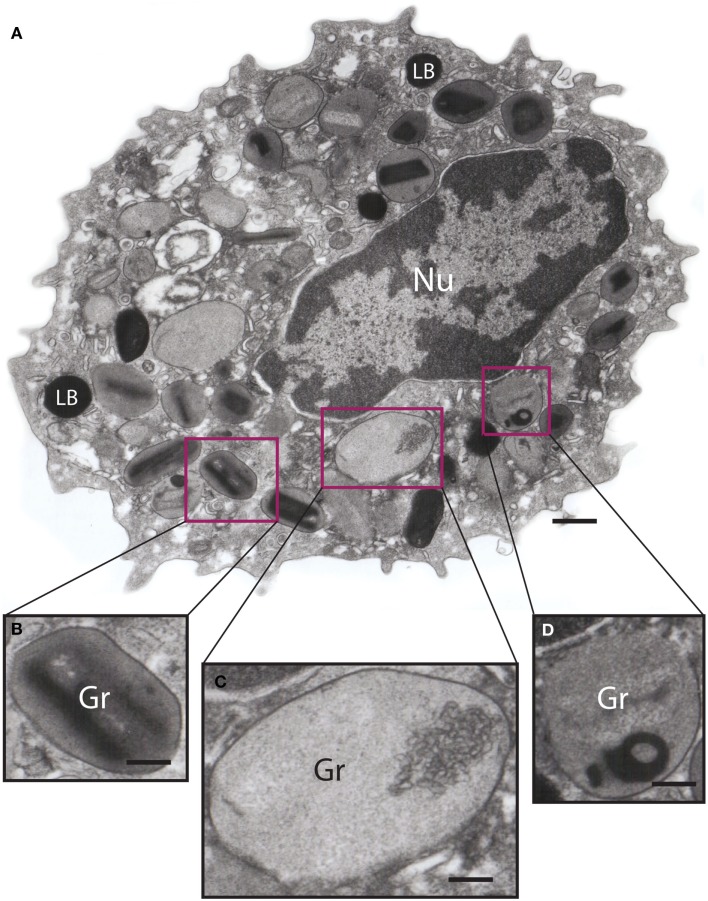
Figure 3.
Ultrastructure of an eotaxin-activated human eosinophil showing piecemeal degranulation (PMD). (A) After stimulation, specific granules (Gr) exhibit different degrees of emptying of their contents and morphological diversity indicative of PMD, such as (B) lucent areas in their cores, (C) enlargement and reduced electron density, and (D) residual cores. Eosinophils were isolated by negative selection from healthy donors, stimulated with eotaxin-1 for 1 h, immediately fixed and prepared for transmission electron microscopy as before (43). Nu, nucleus; LB, lipid body. Scale bar: 500 nm (A); 170 nm (B–D).

