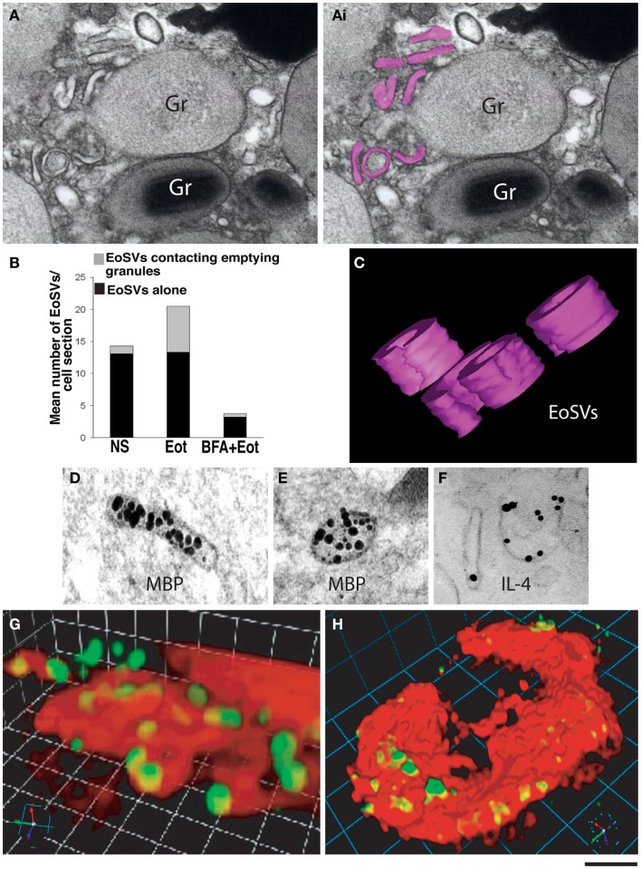
Figure 4.
Vesicular trafficking of granule-derived products from human eosinophils. (A) Eosinophil sombrero vesicles – EoSVs – [highlighted in pink in (Ai)] are observed in the cytoplasm surrounding an emptying, enlarged secretory granule (Gr). An intact granule (Gr) with typical morphology is also observed. (B) Quantification of EoSV numbers revealed significant formation of these vesicles and association with granules undergoing release of their products, after eotaxin-1 (EOT) stimulation (45). Brefeldin-A (BFA) pretreatment suppressed all EoSV numbers dramatically (P < 0.05). NS, not stimulated. (C) Three-dimensional (3D) models obtained from electron tomographic analyses show EoSVs as curved tubular and open structures surrounding a cytoplasmic center. (D–F) As demonstrated by immunonanogold electron microscopy, major basic protein (MBP) (D,E) is transported within the EoSVs lumen, while IL-4 mobilization is associated with vesicle membrane (F). In (G,H), human blood eosinophils suspended in an anti-IL-4 capture antibody-containing agarose matrix were stimulated with eotaxin-1. 3D reconstructed images demonstrate released and captured IL-4 as focal fluorescent green spots at the outer surface of the cell membrane (stained in red). (B,F–H) were reprinted from Ref. (45) and (C–E) from Ref. (46) with permission. Scale bar: 250 nm (A); 150 nm (C–F); 4 μm (G); 6 μm (H).

