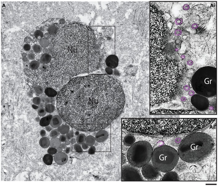
Figure 5.
Ultrastructure of a tissue human eosinophil undergoing cytolysis. Note the disintegrating nucleus (Nu) and extracellular free secretory granules (Gr) in the surrounding tissue. (Ai, Aii) are boxed areas of (A) seen at higher magnification. Note the presence of free, intact eosinophil sombrero vesicles (EoSVs – highlighted in pink) in the tissue, after cell lysis. Tissue eosinophils were present in a biopsy performed on a patient with inflammatory bowel disease. Scale bar: 800 nm (A); 300 nm (Ai, Aii).

