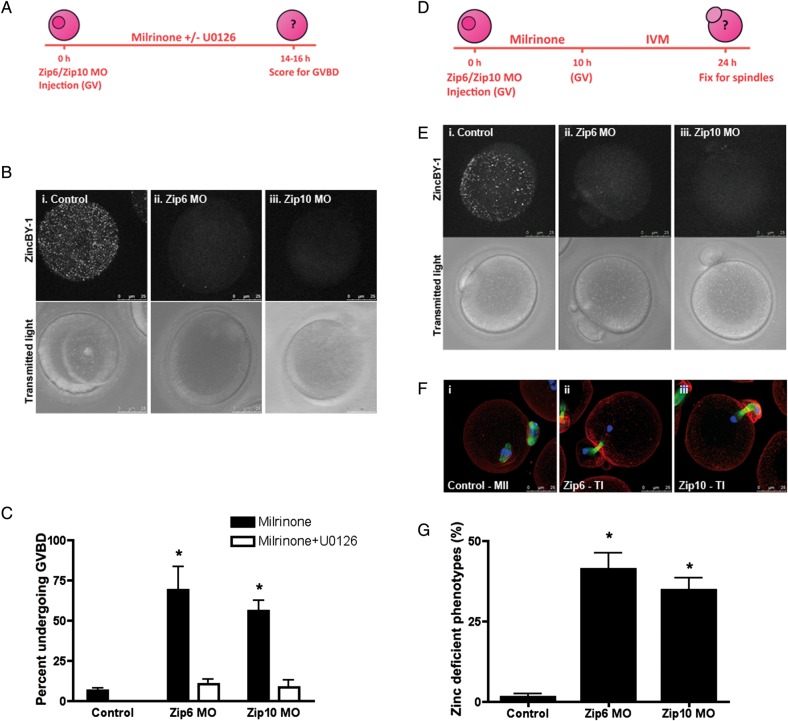
Figure 4.
Disruption of Zip6 and Zip10 expression recapitulate phenotypes of TPEN-induced zinc insufficiency. (A) Oocytes were injected with 5 mM Zip6 or Zip10 morpholino (MO) and held in milrinone medium alone or milrinone medium supplemented with 10 μM U0126. Control oocytes were uninjected and held in milrinone medium. (B) Labile zinc distribution was visualized in injected oocytes by staining with 50 nM ZincBY-1. (C) At the end of culture, the percentage of oocytes undergoing germinal vesicle breakdown (GVBD) was calculated. Black bars represent oocytes held in milrinone, while white bars represent oocytes held in milrinone with U0126. (D–F) Oocytes were injected with 5 mM Zip6 or Zip10 MO, held for 10 h in milrinone medium and then transferred to in vitro maturation medium for 14 h before being imaged for labile zinc distribution (E) or fixed for immunostaining (F). (F) Spindle structure was interrogated by labeling oocytes with α-tubulin (green), actin (red) and chromatin (blue). Representative confocal images of MII and telophase I-like spindles are shown as Z-stack projections. (G) The percentage of oocytes arresting in a telophase I-like phenotype was calculated for control uninjected and Zip6 and Zip10 MO injected oocytes. Graphical data are presented as the mean ± SEM of three independent trials with at least 30 oocytes per trial. Statistical differences were calculated according to one-way ANOVA with Bonferroni post hoc test in comparison to the Control group (P < 0.001). Scale bar, 25 μm.

