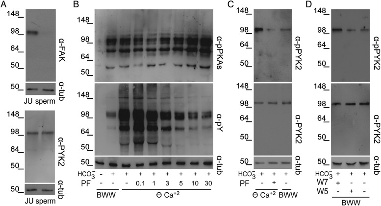
Figure 4.
Presence of PYK2 in human sperm and its activation in ⊖ Ca2+. (A) Jurkat cells (JU), as a positive control for FAK and PYK2 expression, or sperm protein extracts were analyzed by western blotting using α-FAK or α-PYK2 antibodies (n = 3). (B) Sperm were incubated for 6 h in ⊖ Ca2+ containing different concentrations of the FAK/PYK2 inhibitor PF431396, and protein extracts were analyzed for both PKA substrate and Tyr phosphorylations by western blotting using α-pPKAs or α-pY antibodies, respectively (n = 4). (C) Sperm were incubated for 6 h in BWW or in ⊖ Ca2+ with or without PF431396 (10 µM). Protein extracts were analyzed by western blotting using α-pPYK2 antibody and the membrane was then stripped and reprobed with α-PYK2 antibody (n = 4). (D) Sperm were incubated for 6 h in BWW with or without W7 (50 µM) or its analog W5 (50 µM) and protein extracts were analyzed by western blotting using α-PYK2 and then α-pPYK2 antibodies (n = 4). β-Tubulin was used as the protein loading control.

