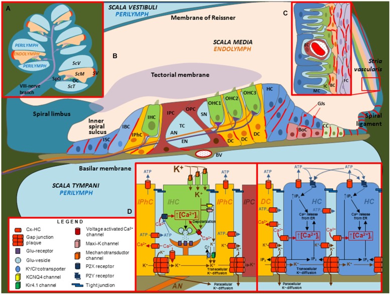
Figure 2.
Illustrations depicting the cellular organization of the mammalian cochlea and the putative roles of Cx channels. (A) Transverse section through the cochlea. OC: organ of Corti. SpG: Spiral ganglia. SV: stria vascularis. ScV: scala vestibuli. ScM: scala media. ScT: scala tympani. (B) Diagram of the organ of Corti. In this representation, the relative sizes of the different structures are altered to emphasize the organ of Corti, which contains inner hair cells (IHC), outer hair cells (OHC), different types of support cells and structures including afferent (AN) and efferent (EN) nerve fibers, blood vessel (BV), Boettcher cells (BoC), Claudius cells (CC), Deiter’s cells (DC), Hensen cells (HC), Inner border cells (IBC), Inner pilar cells (IPC), Inner phalangeal cells (IPhC), Inner Sulcus cells (ISC), Outer pilar cells (OPC), space of Nuel (SN) and tunnel of Corti (TC). Inset (C) Structure of the Stria vascularis containing endothelial cells (EC), fibrocytes (FC), basal cells (BC), intermediate cells (IC) and marginal cells (MC). Inset (D) Putative roles of Cx channels in support cells. Cx HCs and GJ channels in support cells have been proposed to mediate the flux of K+, ATP, IP3 and Ca2+ across the membrane and between cells, respectively.

