Figure 3.

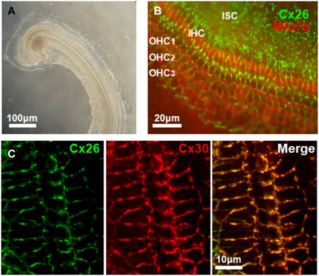
Connexin expression in mouse cochlea. (A) Low-power transmitted-light image of a primary explant culture of cochlea (72 h in vitro) prepared from a P4 mouse. (B) Immunoreactivity for Cx26 (green), and MyosinVIIa (red), marker of hair cells, obtained from an in vitro primary culture of mouse cochlea after 4 days in culture. Cx26 staining is widespread in cochlea support cells in regions of cell-cell contact. Inner (IHC) and outer (OHC) hair cells are indicated. ISC: Inner support cells. (C) Expression of Cx26 and Cx30 in support cells of organ of Corti in an in vitro primary culture of mouse cochlea after 5 days in culture. Merged image of Cx26 and Cx30 immunostaining shows extensive co-localization of both connexins.
