Fig. 2.

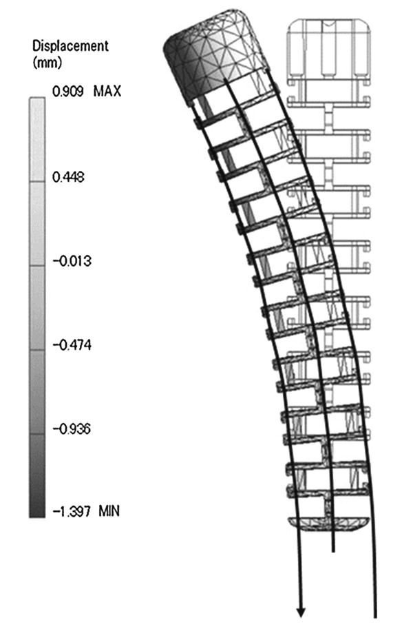
Screen capture during the structural analysis of the catheter CAD model. The displacement of the model is described as in the gray-scale shading map. The bending angle was calculated with the displacement values

Screen capture during the structural analysis of the catheter CAD model. The displacement of the model is described as in the gray-scale shading map. The bending angle was calculated with the displacement values