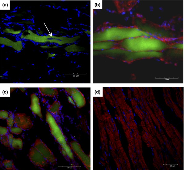
Figure 5.

Presence of GFP+ cells in skeletal muscle of chagasic chimeric mice. Skeletal muscle of chimeric mice euthanized at different time points after infection with T. cruzi was analysed by immunofluorescence microscopy. (a) Parasite nest within a GFP+ myofibre (arrow) after 33 days of infection. (b and c) GFP+ (green) myosin+ (red) myofibres were found in skeletal muscle sections obtained from mice after 60 (B) and 192 (c) days of infection. (d) Skeletal muscle section obtained from an uninfected chimeric mouse. Sections were stained with anti-myosin antibody (red) and DAPI (blue).
