Figure 6.

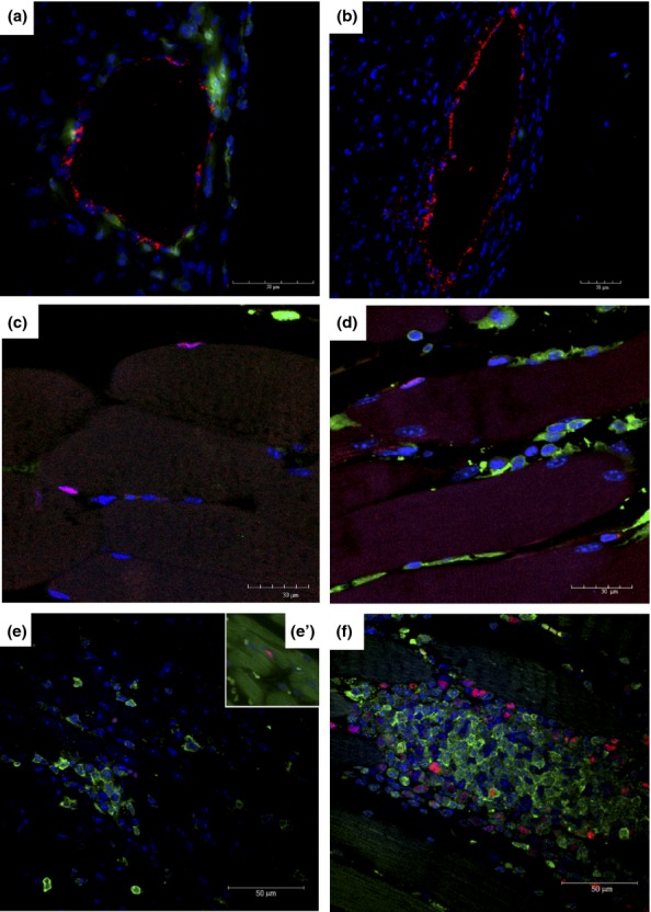
Characterization of GFP+ cells in different organs of T. cruzi-infected chimeric mice. GFP+ cells (green) were observed to be associated with blood vessels in the hearts of mice 33 days after infection (a), but not in uninfected chimeric mice (b). In red, staining for von Willebrand factor. Satellite cells Pax7+ in skeletal muscle sections of naïve (c) and T. cruzi-infected mice (d) 33 days after infection. Presence of GFP+ (green) proliferating PCNA+ cells (red) in the inflammatory infiltrates of the heart (e) and PCNA+GFP− cardiomyocytes stained with phalloidin (green; e’) and skeletal muscle (f) tissue, 33 days postinfection. Nuclei were stained with DAPI (blue).
