Fig. 3.
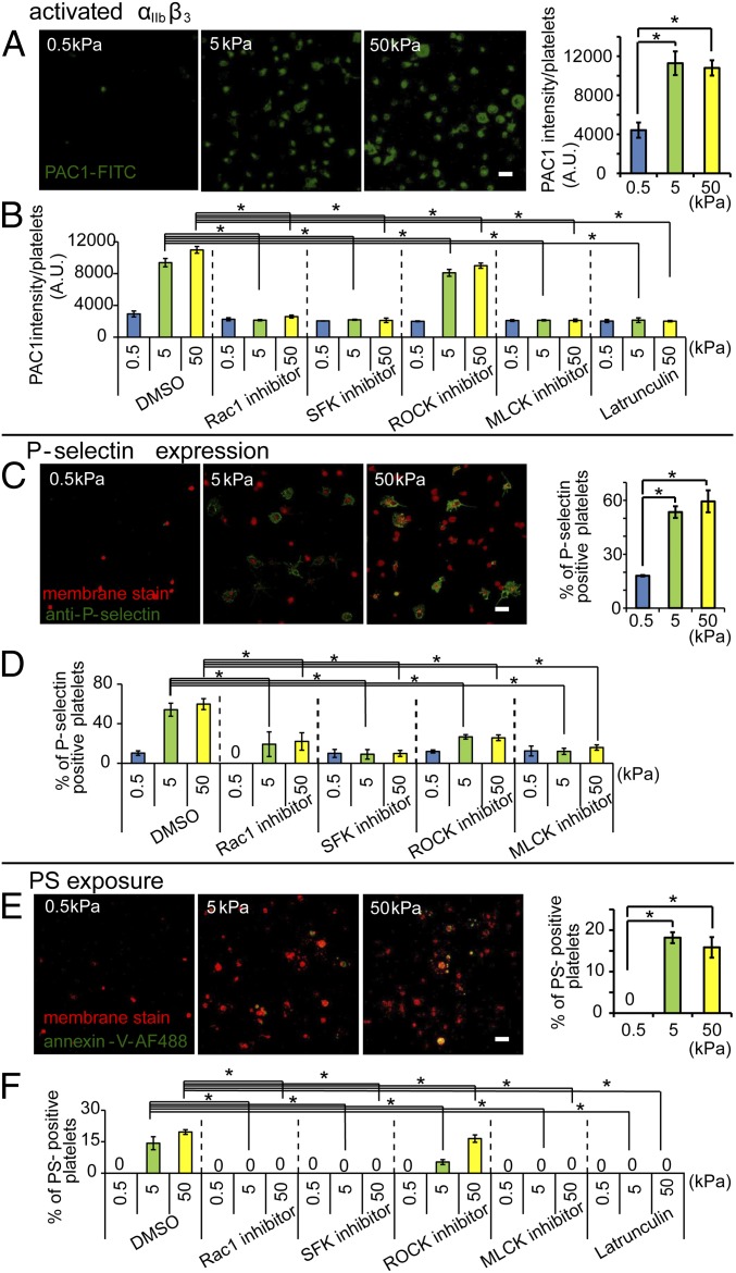
Platelet activation is mediated by substrate stiffness. (A) Increased substrate stiffness increases activation of the αIIbβ3 platelet integrin as measured by PAC-1–FITC immunostaining via confocal microscopy. When normalized to spread platelet surface area, the average intensity is higher for the fibrinogen-conjugated 5- and 50-kPa gels compared with 0.5-kPa gels. (B) Inhibition of Rac1, SFK, MLCK, and actin polymerization attenuates the substrate stiffness-mediated αIIbβ3 activation of adherent platelets as measured by the average intensity of PAC-1–FITC staining. (C) Increased substrate stiffness increases P-selectin expression on adherent platelets as measured by P-selectin immunostaining (red: fluorescent membrane dye; green: P-selectin immunostaining) via confocal microscopy. (D) Inhibition of Rac1, SFK, ROCK, and MLCK differentially attenuate the substrate stiffness-mediated P-selectin expression on adherent platelets. (E) PS exposure was found only on platelets adhered onto stiffer gels of 5 and 50 kPa as measured as measured by Annexin V staining via confocal microscopy. (F) Inhibition of Rac1, SFK, MLCK, and actin polymerization abolished the substrate stiffness-mediated PS exposure on adherent platelets. (P < 0.05; n = 3 experiments; error bars indicate SD.) (Scale bar: 10 μm.)

