Fig. 4.
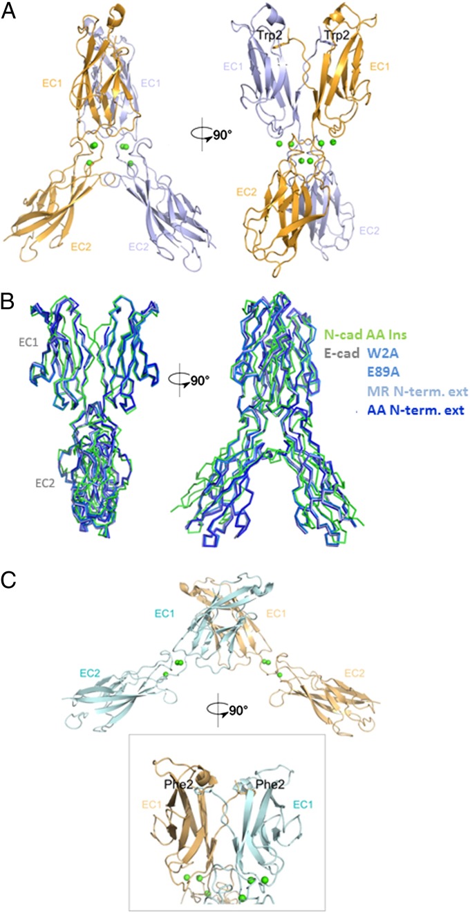
Structure of N-cadherin W2F and N-cadherin AA-insertion mutants. (A) Structure of EC1–EC2 dimer of N-cadherin AA-insertion mutant. The two protomers are shown in light blue and bright orange, with the exchanged Trp2 side-chain shown. Calcium ions are shown as green spheres. (B) Ribbon representation showing the superposition of the N-cadherin AA-insertion mutant (green) and known E-cadherin X-dimer mutants (shades of blue): E-cadherin W2A (PDB ID code 3LNH), E89A (PDB ID code 3LNI), N-terminal MR-extension (PDB ID code 1EDH), and N-terminal AA-extension (PDB ID code 3LNG). (C) Structure of EC1–EC2 dimer of N-cadherin W2F mutant. Protomers are shown in pale cyan and light orange, calcium ions in green spheres. A close-up of the swapped interface, with the exchanged Phe2 side chains, is shown.

