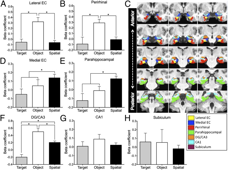
Fig. 2.
Region of interest responses during correct rejections of object and spatial lures. (A and B) Increased activity during object discrimination trials in LEC and PRC. (C) ROI segmentation on average group template in the coronal view. Representative slices are arranged from left to right then top to bottom in the anterior-posterior direction, and ROI demarcations are represented in accordance with the color key displayed below. (D and E) Increased activity during spatial discrimination trials in MEC and PHC. (F–H) Increased activity during both object and spatial discrimination trials in DG/CA3 but not in CA1 and subiculum. Baseline (β coefficient = 0) was correct rejection of novel foil images. DG, dentate gyrus; EC, entohrinal cortex. Asterisks indicate significance at P < 0.05 corrected.

