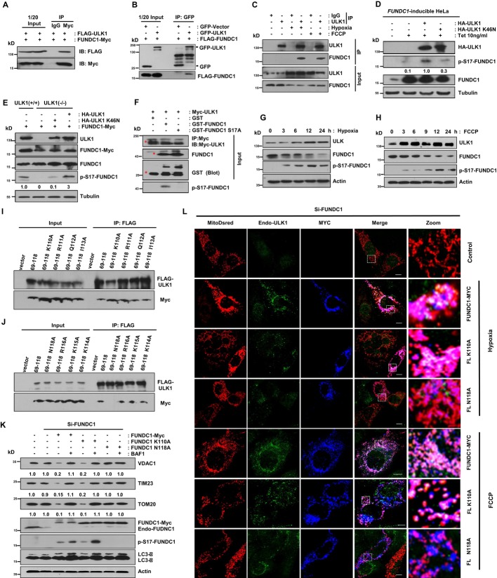
Figure 2. The mitophagy receptor FUNDC1 is a novel substrate of ULK1 and FUNDC1 (N118A) that prevents its binding to ULK1, impairs ULK1 translocation to mitochondria, and inhibits mitophagy.
A HeLa cells were co-transfected with FUNDC1-Myc and FLAG-ULK1. Cells were lysed and immunoprecipitated with anti-Myc antibody 24 h after transfection.
B FLAG-FUNDC1 was co-transfected with GFP-vector or GFP-ULK1 for 24 h in HeLa cells, and then, cell lysates were immunoprecipitated with anti-GFP antibody.
C MEFs were exposed to hypoxia (1% O2) for 12 h or FCCP (20 μM) for 3 h, followed by immunoprecipitation using anti-ULK1 antibody.
D The expression of FUNDC1 is induced by 10 ng/ml tetracycline (Tet) in FUNDC1-inducible HeLa cells. Cells were transfected with HA-ULK1 or HA-ULK1 (K46N) for 24 h. Then, the cell lysates were prepared for immunoblotting using indicated antibodies.
E ULK1+/+ cells were transfected with FUNDC1-Myc, and ULK1 cells were transfected with FUNDC1-Myc alone, FUNDC1-Myc and HA-ULK1, or FUNDC1-Myc and HA-ULK1 (K46N). Cells were harvested and lysed for immunoblots 24 h after transfection.
F Purified GST-FUNDC1 and GST-FUNDC1 (S17A) were subjected to an in vitro kinase assay with Myc-ULK1 immunoprecipitated by anti-Myc antibody from Myc-ULK1-transfected cells about 24 h post-transfection. Phosphorylated FUNDC1 was detected by anti-p-S17-FUNDC1 antibody. The red asterisks mark the target bands.
G Western blot analysis of the kinetics of ULK1, FUNDC1, and the phosphorylated FUNDC1 in MEFs exposed to hypoxia (1% O2) for the indicated times.
H Western blot analysis of the kinetics of ULK1, FUNDC1, and the phosphorylated FUNDC1 in MEFs treated with FCCP (20 μM) for the indicated times.
I, J The single mutant which can abolish the ULK1 and FUNDC1 interaction was identified. HeLa cells were co-transfected with Flag-ULK1 and the indicated constructs. 24 h after transfection, cells were lysed for immunoprecipitation by anti-Flag antibody.
K MEFs were transfected with FUNDC1-Myc and its mutants after the endogenous FUNDC1 was knocked down by siRNA. 36 h after transfection, cells were harvested in the absence or presence of 50 nM bafilomycin A1 (BAF1) and then lysed for immunoblotting with the indicated antibodies.
L MEFs were co-transfected with FUNDC1-Myc or its mutants and MitoDsred after the endogenous FUNDC1 was knocked down by siRNA. 24 h post-transfection, cells were treated with hypoxic (1% O2) conditions for 12 h or FCCP (20 μM) for 6 h before fixed by 4% paraformaldehyde and stained with indicated antibodies. Scale bar, 10 μm.
Data information: All results are from three independent experiments.
Source data are available online for this figure.

