
Figure 1. PICK1 interacts with Ago2.
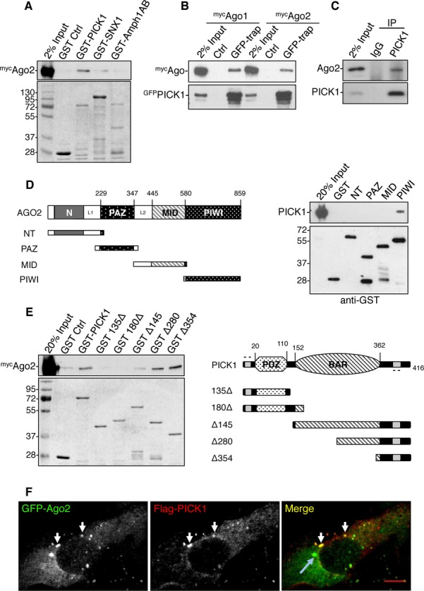
A Ago2 binds to GST-PICK1 but not other BAR-domain proteins tested. GST-PICK1, GST-SNX1 (sorting nexin 1), and GST-Amph1AB (Amphiphysin1 domains AB) were incubated with HEK293 cell lysate expressing mycAgo2. Bound proteins were detected by western blotting using anti-myc or by Coomassie staining.
B GFP-PICK1 interacts with both mycAgo1 and mycAgo2 from HEK293 cells. Lysates were incubated with GFP-trap agarose or blocked agarose beads as control, and bound proteins were detected by western blotting using anti-myc or anti-GFP.
C Ago2 interacts with PICK1 in neurons. Lysates were immunoprecipitated with anti-PICK1 or control IgG, and bound proteins were detected by western blotting.
D PICK1 interacts with the PIWI domain of Ago2. GST fusions of truncation mutants for the N-terminus (GST-NT) or the PAZ, MID, or PIWI domains as depicted in the diagram were incubated with purified his6-PICK1. Bound protein was detected by western blotting.
E Ago2 interacts with the C-terminus of PICK1. GST-PICK1 full-length or truncation mutants as depicted in the diagram were incubated with HEK293 cell lysate expressing mycAgo2. Bound proteins were detected by western blotting with anti-myc or by Coomassie staining.
F PICK1 colocalizes with Ago2 puncta distinct from P-bodies. COS7 cells expressing GFP-Ago2 and flag-PICK1 were stained with anti-flag. White arrows indicate overlapping puncta between Ago2 and PICK1; blue arrow indicates a P-body; scale bar, 10 μm.