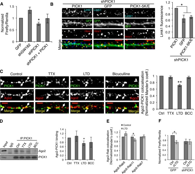
Figure 4. PICK1 is involved in microRNA-mediated silencing pathways during chemical LTD.
A PICK1 knockdown causes an increase in translational repression in cultured neurons. Dual-luciferase assay was performed in neurons expressing Renilla and Firefly luciferase reporter containing Limk1 3′UTR and either GFP, shGW182, shPICK1, or shPICK1 + sh-resistant GFP-PICK1. Values were normalized to the GFP-alone condition. *P < 0.05 (Student’s t-test with Bonferroni correction), n = 4–7.
B PICK1 knockdown causes a decrease in endogenous Limk1 expression in neuronal dendrites. Neurons expressing shPICK1 plus GFP, sh-resistant GFP-PICK wild-type, or sh-resistant GFP-PICK 5K/E mutant were stained for Limk1 (blue) and the synaptic marker Homer1 (red). Scale bars, 5 μm. Graph shows Limk1 immunofluorescence normalized to GFP-PICK1 wild-type rescue condition. *P < 0.05 (Student’s t-test with Bonferroni correction), n > 14 cells per condition.
C Chemical LTD causes a decrease in colocalization between Ago2 and PICK1 in dendrites of hippocampal neurons. Hippocampal neurons were treated with tetrodotoxin (TTX), Bicuculline (BCC), or NMDA (LTD), as shown and stained for PICK1 (green) and Ago2 (red). Arrows indicate overlapping puncta positive for PICK1 and Ago2. Scale bars, 5 μm. Graph shows Mander’s coefficients for the fraction of Ago2 colocalized with PICK1, normalized to untreated controls. **P < 0.01 (Student’s t-test with Bonferroni correction), n > 16 cells per condition.
D Chemical LTD reduces the Ago2–PICK1 interaction. Neuronal cultures were treated as in (C), lysates were immunoprecipitated with anti-PICK1 or control IgG and bound proteins detected by western blotting. Values for Ago2 were normalized to PICK1 IP and to untreated control. *P < 0.05 (Student’s t-test with Bonferroni correction), n = 5–7.
E Chemical LTD reduces the colocalization between Ago2 and Rab11. Neurons were treated for chemical LTD and stained with Ago2 and Rab proteins. Representative images are shown in Supplementary Fig S2B. Graph shows Mander’s coefficients for the fraction of Ago2 colocalized with Rab protein, normalized to untreated controls. *P = 0.03 (Student’s t-test), n > 9 cells per condition.
F PICK1 knockdown occludes the effect of chemical LTD on translational repression. Dual-luciferase assay was performed in cultured neurons expressing GFP or GFP + shPICK1 and luciferase constructs as in (C). Neurons were treated for chemical LTD and lysed after 5 min. Relative luciferase values were normalized to vehicle-treated GFP control. *P = 0.03 (Student’s t-test), n = 8.
Source data are available online for this figure.

