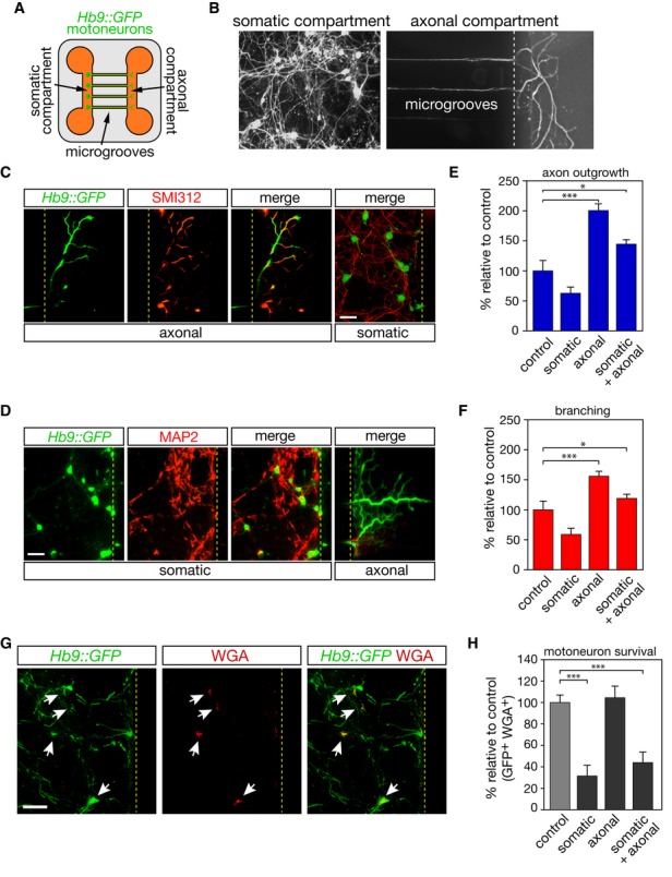
Figure 3. Somatic or axonal LIGHT signaling elicits opposite responses in motoneurons.

A Schematic representation of the microfluidic device.
B Motoneurons are plated into the somatic compartment and they extend their axons into the axonal compartment through 750-μm-long microgrooves.
C, D Hb9::GFP motoneurons were cultured for 120 h and immunostained using SMI312 (C) and MAP2(D). Scale bar, 50 μm.
E, F After 120 h of culture, sLIGHT (100 ng/ml) was added either in the somatic or in the axonal compartment, or both. Axonal length (E) and branching (F) were determined 48 h later.
G The retrograde transport of Alexa555-conjugated WGA allows tracing of motoneurons whose axons have crossed microchannels (white arrows).
H The survival of motoneurons whose axons have reached the axonal compartment is determined 48 h later by counting the number of GFP- and WGA-positive cells. Scale bar, 100 μm.
Data information: Histograms show mean values ± SEM in three independent experiments, ANOVA with Tukey–Kramer’s post hoc test; *P < 0.05, ***P < 0.001
