
Figure 5. LIGHT is required for axonal regeneration following nerve lesion.
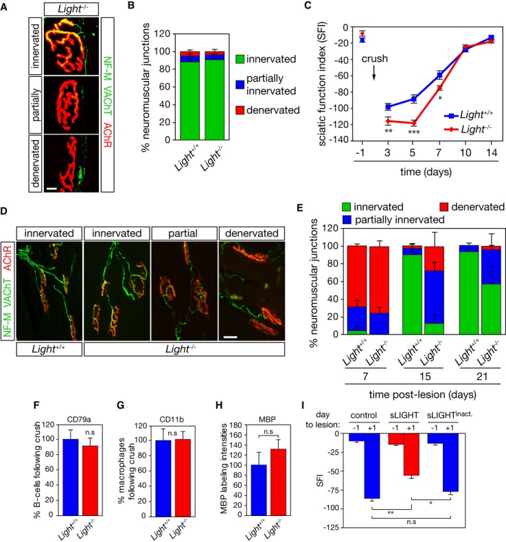
A Tibialis anterior muscle from 50-day-old Light+/+ (not shown) Light−/− mice was stained with anti-neurofilament (NF-M) and anti-vesicular acetylcholine transporter (VAChT) antibodies (in green) and fluorochrome-conjugated α-bungarotoxin (AChR, in red). Scale bar, 10 μm.
B The percentage of innervated, partially innervated and denervated motor endplates, based on the overlap of NF-M/VAChT and AChR area, was quantified (n = 3).
C Functional recovery was quantified by calculating the SFI before and on days 3, 5, 7, 10, and 14 post-nerve lesion in Light+/+ (n = 24) and Light−/− mice (n = 22).
D Representative confocal images illustrating complete to partial occupancy and vacancy of neuromuscular junctions following nerve lesion. Scale bar, 50 μm.
E The degree of neuromuscular junction occupancy was quantified after nerve injury in Light+/+ and Light−/− mice (n = 4).
F–H The number of CD79a+ (F), CD11b+ (G) and the intensity of MBP (H) immunoreactivity were quantified in the sciatic nerve of Light+/+ and Light−/− mice 10 days after crush (n = 3).
I At the time of a unilateral sciatic nerve lesion, mice were intramuscularly injected with PBS (control), sLIGHT and a heat-inactivated sLIGHT (250 ng/ml). The SFI was determined at indicated days to the lesion (n = 12 for each condition).
Data information: *P < 0.05, **P < 0.01, ***P < 0.001. In (C, I) two-way repeated-measure ANOVA, Newman–Keuls post hoc test; in (F, G, H) unpaired two-tailed Student’s t-test.