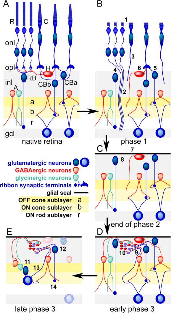
Figure 1.

Cellular composition of the retina and the three phases of remodeling associated with retinal degenerations. (Panel A) The laminated retina with rod (R) and cone (C) photoreceptor cells in the outer nuclear layer (onl) and synaptic endings in the outer plexiform layer (opl) driving rod bipolar cells (RB), ON (CBb) or OFF (CBa) cone bipolar cells, and/or horizontal cells (H). Rod and cone bipolar cells then drive separate sets of GABAergic (red) and glycinergic (green) amacrine cells (A) in the OFF (a), ON (b), and rod (r) layers of the inner plexiform layer. Only cone bipolar cells drive ganglion cells that project to the brain through the optic nerve. The ganglion cell layer (gcl) contains the cell bodies of ganglion cells positioned next to the vitreous of the eye. It is the first layer accessible through surgical approaches or intraocular injections. (Panel B) Phase 1. Stressed photoreceptor cells truncate their outer segments (1) and extend anomalous axons deep into the retina (2,3). Bipolar cells retract dendrites (4,5) and horizontal cells generate ectopic axons. (Panel C) End of phase 2. Death of photoreceptor cells and ablation of the outer nuclear layer. The remnant retina is capped by a seal of glial processes (7) and all bipolar cells have truncated their dendrites. (Panel D) Early phase 3. All surviving cells can initiate neuritogenesis, forming ectopic fascicles of processes (9) and new synaptic microneuromas (10). (Panel E) Late phase 3. Cells revise their connection repertoires (11) and some migrate to ectopic sites (12), ganglion cells generate new intraretinal axons (13), and rod bipolar cells decrease their synaptic ribbon size (14). Neuronal cell death progresses (stippled cells).
