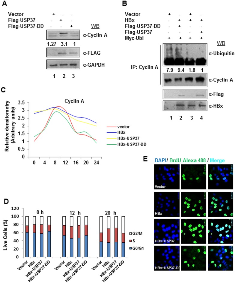
Figure 1. USP37 and HBx act in synergy to deregulate cell Cycle.
(A) Huh7 cells were transfected with Vector, Flag-USP37 and Flag-USP37-DD construct and the levels of USP37 protein were measured by western blotting (WB). (B) Ubiquitination assay was performed with lysates from cells transiently expressing Vector, HA-HBx, Flag-USP37, Flag-USP37-DD and Myc-Ubi as indicated (Cells in 100 mm dish were transfected with 4 µg Vector and 2 µg Myc-ubiquitin; 2 µg Vector, 2 µg HA-HBx and 2 µg Myc-ubiquitin; 2 µg Flag-USP37, 2 µg HA-HBx and 2 µg Myc-ubiquitin or 2 µg Flag-USP37-DD, 2 µg HA-HBx and 2 µg Myc-ubiquitin) and treated with 20 µM MG132 for 6 h, by immunoprecipitating Cyclin A. Immuno-complexes were eluted and western blotted with α-Ubiquitin antibody. (C) Cyclin A expression was chased in IHH cells transiently transfected with Vector, HA-HBx, Flag-USP37 and Flag-USP37-DD as indicated and harvested at indicated time intervals post 72 hrs serum starvation. (D) IHH cells transfected with Vector control, HA-HBx, Flag-USP37 and Flag-USP37-DD as indicated, were synchronized in G0/G1 phase by Serum starvation followed by harvesting at indicated time points. Cells in different phases of cell cycle were analyzed by flow cytometry. Values are represented as bar diagrams (E) Brd-U incorporation assay was carried out in Huh7 cells transfected with Vector control, HA-HBx, Flag-USP37 and Flag-USP37-DD as indicated, by incorporating BrdU followed by staining with antibody against BrdU and counterstaining with DAPI to observe actively replicating cells as seen in the representative confocal images. Scale bar represents 50 µm.

