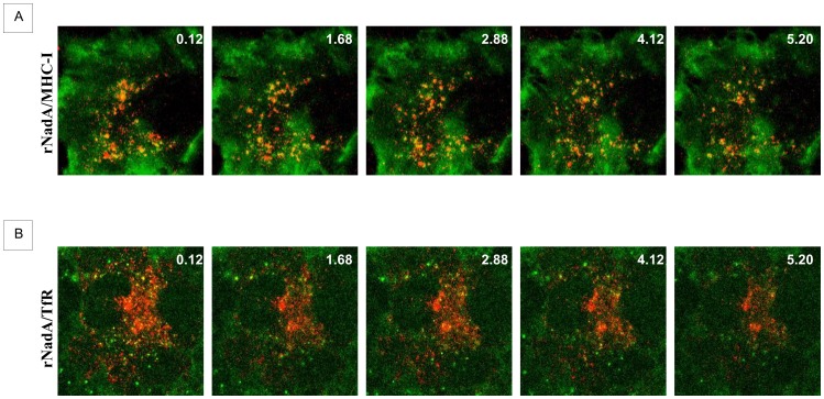
Figure 6. Time lapse rNadA internalization.
A: Chang cells were incubated for 1 h with Alexa633-conjugated rNadA (shown in red) and Alexa488-conjugated anti-MHC-I antibody (green). Cells were then washed and live images were recorded every 2 seconds by confocal microscopy. Five frames from the Movie S1 are shown. Time (seconds) is indicated in the top right of each panel. B: Chang cells were incubated for 1 h with Alexa488-conjugated rNadA (green) and Cy3-conjugated transferrin (red). Cells were then washed and live images were recorded every 4 seconds by confocal microscope. Five frames from the Supplementary Movie S2 are shown. Time (seconds) is indicated in the top right of each panel.

