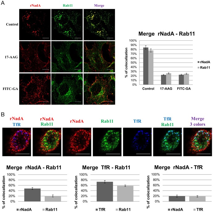
Figure 9. rNada internalization and colocalization with Rab11 and TfR.
A: rNadA and Rab11 colocalization in presence of inhibitors. The control untreated Chang cells are shown in the upper panel. Pre-treatment of cells was performed for 1 hour with 10 µM 17-AAG (middle panel) or 10 µM FITC-GA (bottom panel). Cells were then incubated with 200 µg/ml rNadA at 37°C for 1 hour. Afterward, cells were fixed, permeabilized and stained for NadA. The drugs were present during the entire incubation period. Graph: rNadA columns indicate the percentage of rNadA immunofluorescent pixels colocalizing with Rab11 immunofluorescent pixels in untreated, 17-AAG and FITC-GA cells (from left to right) respectively. Conversely, Rab11 columns indicate the percentage of Rab11 immunofluorescent pixels colocalizing with rNadA immunofluorescent pixels in untreated, 17-AAG and FITC-GA cells (from left to right) respectively. B: Rab11, rNadA and TfR colocalization . A: Chang cells were transfected with EGFP-Rab11 plasmid, incubated 24 hours at 37°C. and then incubated with 200 µg/ml of rNadA at 37°C for 2 hours. Afterward, cells were fixed, permeabilized and stained for NadA (Red) and TfR (blu). Rab11 is colored in green. Graphs: rNadA columns indicate the percentage of rNadA immunofluorescent pixels colocalizing with Rab11 fluorescent pixels (left) or with TfR immunofluorescent pixels (right). Rab11column indicate the percentage of fluorescent pixels colocalizing with rNadA immunofluorescent pixels (left) or TfR immunofluorescent pixels (right). TfR columns indicate the percentage of TfR immunofluorescent pixels colocalizing with Rab11 fluorescent pixels (middle) or with rNadA immunofluorescent pixels (left). Data are mean ± s.e.m representative of two independent experiments, each assessing 20–25 cells.

