Figure 5.
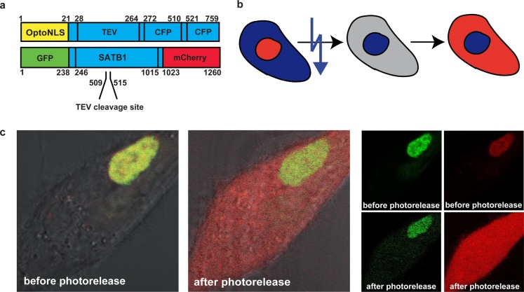
Photocontrol of intranuclear SATB1 cleavage. (a) Photocontrolled delivery of the TEV protease into the nucleus of MCF10A cells and subsequent specific cleavage of a transcription factor. The TEV protease has a prepended OptoNLS and two attached CFPs, to block passive diffusion into the nucleus. A TEV protease site was engineered into the SATB1 transcription factor, which was also flanked by two fluorophores to allow intranuclear cleavage to be visualized. (b) TEV-protease (blue) is kept in the cytoplasm and enters the nucleus after photoactivation. SATB1 in the nucleus (yellow) containing a TEV target site will remain intact until the protease enters the nucleus and starts cleavage. Upon cleavage of the protein, its part containing the NLS will remain nuclear whereas the other part (red) will distribute all over the cell. (c) Fluorescence images: before photorelease of the TEV protease, GFP (green), and mCherry (red) are colocalized and fully contained in the nucleus (left panel), confirming that an intact SATB1 is expressed and localized in the nucleus. After photorelease of TEV, it enters the nucleus and there it cleaves SATB1. The green N-terminal fragment stays in the nucleus since it has a functional NLS; the red C-terminal fragment transitions to the cytoplasm (middle). (right) Images split into single color channels.

