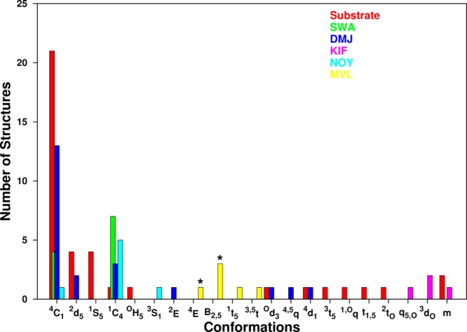
Figure 6.
Comparison of ring conformations of mannopyranose substrates (red) extracted from cocrystal structures with mannosidases (n = 41), and the inhibitors (n = 50) swainsonine (SWA, green), deoxymannojirimycin (DMJ, blue), kifunensine (KIF, pink), noeuromycine (NOY, cyan), and mannoimidazole (MVL, yellow). Presumed transition states for mannosidases indicated by (*).

