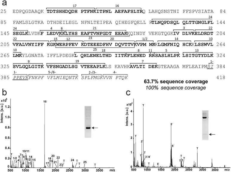
Figure 4. Peptide mapping of EspPα cleavage products of α1-PI.
α1-PI fragments were subjected to tryptic in-gel-digest and generated peptides were analyzed via MALDI-TOF-MS. a, Sequence coverage of α1-PI fragments. Peptides of the large fragment are given in bold and numbered 1–25. Peptides of the small fragment are given in italics and numbered 1′-6′. Note the newly formed N-terminus of the small fragment (SIPPEVK, underlined). b, MALDI-TOF-MS spectrum of the large fragment of α1-PI. Inset: SDS-PAGE gel, glycine buffer. Fragment used for peptide mapping is marked by arrow. c, MALDI-TOF-MS spectrum of the small fragment of α1-PI. Inset: SDS-PAGE gel, tricine buffer. Fragments used for peptide mapping are marked by arrow. α1-PI peptides are numbered according to a, T, trypsin autoproteolysis products, E, EspPα autoproteolysis products.

