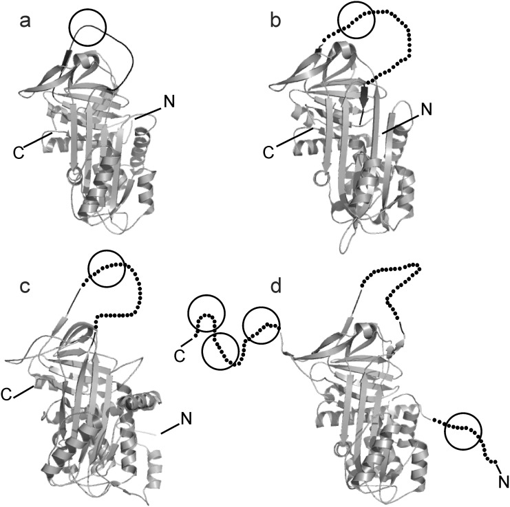
Figure 7. Crystal structures of serpins cleaved by EspPα.
Serpins are shown as cartoons. RCL is indicated in black, approximate cleavage sites are encircled. Non-resolved parts of the crystal structures are indicated by dots (c, RCL of AGT, d, RCL of α2-AP and the N- and C-terminal extension of α2-AP). a, human α1-PI, b, cleaved human α1-AC, the RCL is indicated by dots, c, human angiotensinogen, d, murine truncated α2-APΔ43, the N-terminal extension of native α2-AP is indicated by dots.

