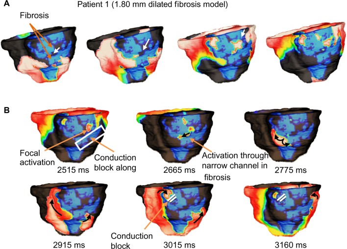
Figure 7.
Sample of VT reentry patterns in Patient 1 model with increased (1.80 mm dilated) fibrosis. (A) Activations are conducted through narrow channels (white arrows) formed within the fibrosis during apex pacing; and (B) The narrow fibrosis channels effectively create a pathway for activations which were conducted along the fibrotic edges. Although figure-of-eight reentry can be observed, the clockwise inferior spiral becomes stifled by conduction block caused by fibrosis. The counter-clockwise inferior/lateral spiral sustains the reentry along fibrosis edge.

