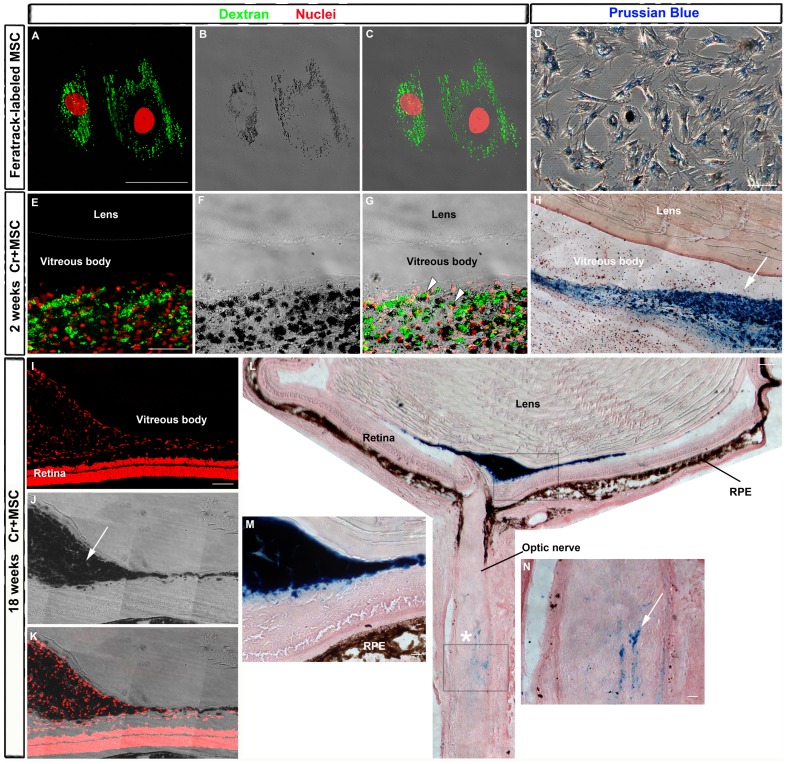
Figure 4. Detection of FeraTrack-labeled MSCs in vitro and after transplantation.
A: Cells were labeled with FeraTrack for 4 h, fixed, and immunostained with anti-dextran antibody (green) to identify dextran-coated particles and nuclei stained with TOPRO (red) (A and C). Iron from SPION is visible as dark spots inside the cell (B-C) or after Prussian blue reaction (D). E-G: Sixteen days after nerve injury and MSC injection, these cells are found in the vitreous body as dextran-positive cells (green) with dark spots (arrowheads in G). Nuclei were stained with TOPRO (red, in E and F). H: Photomontage of transmitted light images of a cryostat section through the eye reacted with Prussian blue. Labeled cells are found mostly in the vitreous body (arrow). I-N: Eighteen weeks after nerve injury and cell transplantation, SPION are visible as dark spots by transmitted light (arrow in J) and also after Prussian blue reaction (L-N). I-K: Photomontage of confocal images from the eye; nuclei stained with TOPRO (red, in I and K). L: Photomontage of transmitted light images of the eye and proximal optic nerve after Prussian blue reaction. Iron was detected mainly in the vitreous body (blue in M), but a small amount was found also in the optic nerve (blue in N), close to the crush site (asterisk). (M,N) Higher magnification of upper (M) and lower (N) boxes in L. RPE, retinal pigmented epithelium. Scale bar: 50 µm.

