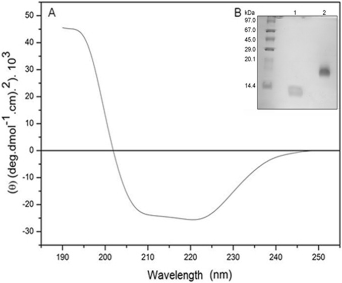
Figure 1. Structural properties of Mo-CBP3.
(A) Circular dichroism spectra (Far-UV) of Mo-CBP3 (2.22 mM) in 20 mM sodium phosphate buffer, pH 7.0, using a rectangular quartz cuvette with a 0.1 cm path length. (B) Denaturing polyacrilamide gel electrophoresis (SDS-PAGE - 15% acrylamide gel) of Mo-CBP3. Molecular mass standards are shown (in kDa) on the left; Lanes 1 and 2, Mo-CBP3 (20 µg) in reducing (4 kDa and 8 kDa subunits) and non-reducing conditions (18 kDa), respectively.

