
Fig. 1.
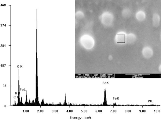
Scanning electron microscopy of the MP-OHP nanocarriers showing spherical morphology of about 100–200 nm size (inset) and the energy dispersive X-ray analysis (EDAX) of a representative nanocarrier marked in the inset revealed co-localization of the characteristic K-x-rays of Fe and Ca as a marker of MNP encapsulated in calcium pectinate.