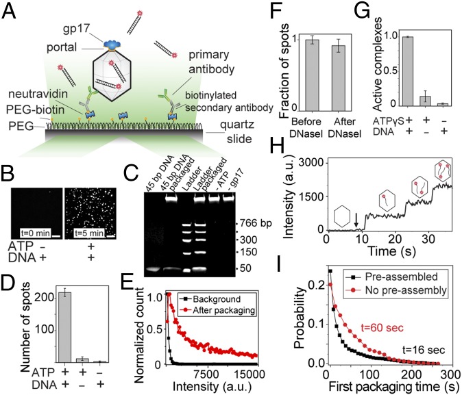
Fig. 1.
Single-molecule fluorescent assay to study DNA packaging initiation. (A) Preassembled phage head–motor (packaging machine) complexes are immobilized on a passivated surface using antibodies against phage T4 capsid proteins. To initiate packaging, fluorescently labeled DNA molecules and ATP are applied and imaged by a total internal reflection microscope. (B) Representative fluorescence images of the slide surface with preassembled packaging complexes and 4 nM DNA before (Left) and 5 min after (Right) introducing 4 nM DNA and 1 mM ATP. (Scale bars, 5 µm.) (C) Bulk packaging assay verifying that the 45-bp Cy5-labeled dsDNA used in the single-molecule assay can be efficiently packaged in bulk (lane 2) and the packaging activity is DNA- and ATP-dependent. Note that about 30% of the ladder DNA added to the reaction (300 µg) was packaged in the control assay (lane 4; compare with 100 ng ladder DNA in lane 3). The high-molecular-weight DNA band in the wells corresponds to the ∼8-kb DNA present in the heads (34). (D) Quantification of the data shown in B. Average number of fluorescent spots per imaging area (70 × 35 µm) in the presence or absence of Cy5-labeled DNA (4 nM) or ATP (1 mM). Error bars represent ± SEM of 30 different imaging areas. (E) Normalized intensity distribution of fluorescent spots before (black) and 10 min after (red) introducing 4 nM DNA and 1 mM ATP. The background spots are significantly dimmer than the spots due to the active packaging. (F) The packaged DNA molecules are protected from DNase I digestion. Number of fluorescent spots 10 min after introducing 4 nM DNA and 1mM ATP followed by DNase I treatment, normalized to the number of spots before DNase I treatment. Error bars represent ± SEM (n = 30). (G) The role of nucleotide or priming DNA in forming active packaging complexes. Fraction of packaging-capable complexes preassembled with or without 1 mM ATPγS or 200 nM unlabeled priming DNA. (H) Fluorescence intensity time trace of a single packaging complex as it packages three Cy5-labeled DNA molecules in succession. The arrow denotes the time when 1 mM ATP and 4 nM DNA were flowed into the chamber. (I) Normalized probability of the first packaging times for preassembled complexes (black) in the presence of 1 mM ATP and 4 nM Cy5-labeled DNA and for de novo assembled complexes (red) in the presence of 1 mM ATP, 4 nM Cy5-labeled DNA, and 2 µM gp17.

