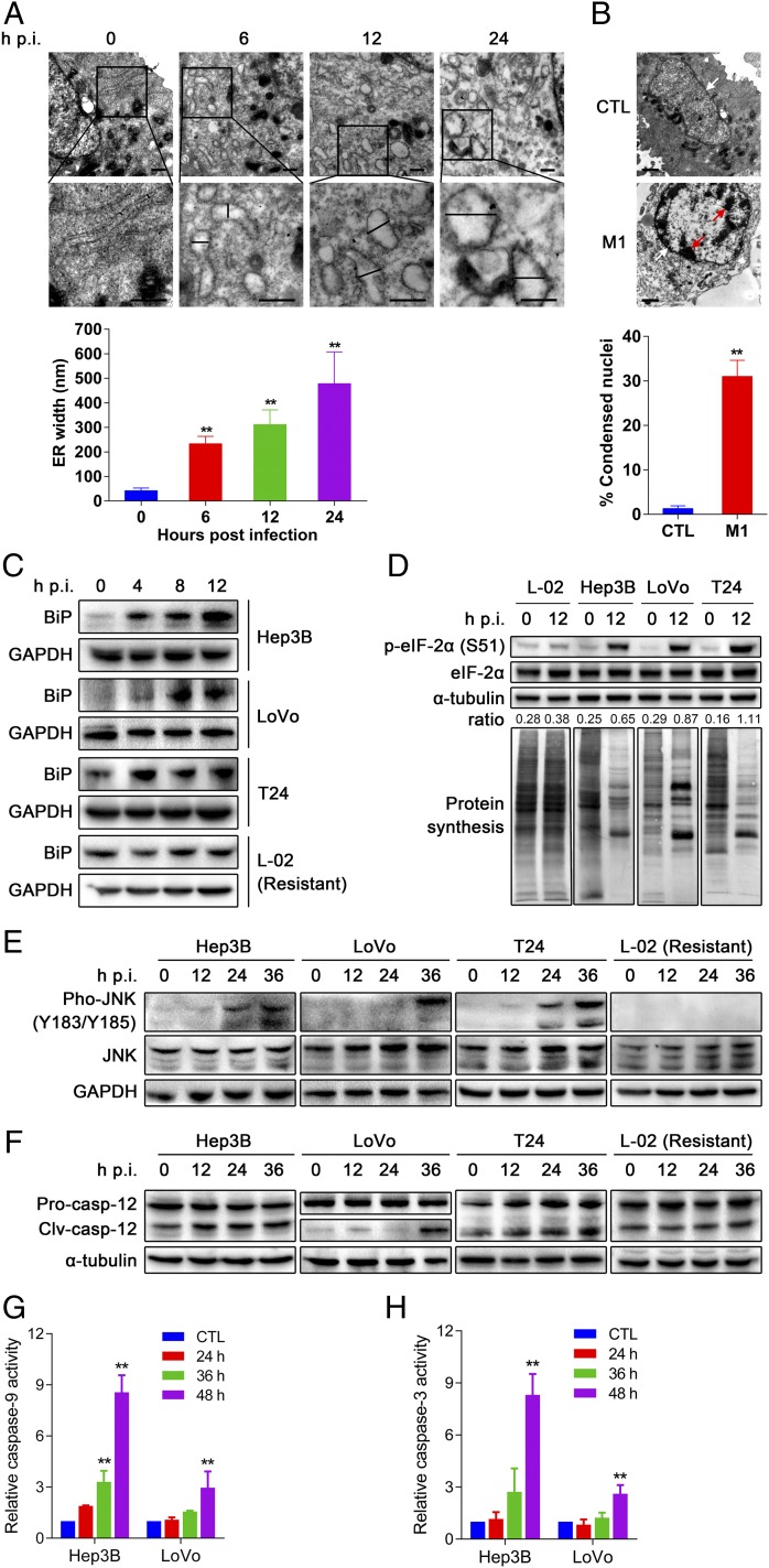
Fig. 2.
M1 triggers prolonged and severe ER stress-mediated apoptosis in susceptible cancer cells. (A) Observation of ER distension in Hep3B cells infected with M1 by transmission EM. Middle shows higher magnification from the box in Top. (Scale bars: 500 nm.) Quantification of ER distension is also presented. (B) Observation of chromatin condensation in infected Hep3B cells by transmission EM. Red arrows, condensed chromatin; white arrows, nuclear envelope. (Scale bars: 1 μm.) Quantification of condensed nuclei is also presented. (A and B) Means ± SDs from three independent experiments are shown. (C–F) The effect of M1 on ER stress signal pathways. Western blot analyses of (C) BiP, (D) phosphorylated eIF-2α (S51), (E) phosphorylated JNK (Y183/Y185), and (F) cleaved casepase-12 (Clv-casp-12) are shown. GAPDH and α-tubulin served as loading controls. The ratio between phosphorylated eIF-2α and α-tubulin was calculated. Pro-casp-12, pro-caspase-12. (D) Detection of protein synthesis after M1 infection (MOI = 10, 12 h) by l-AHA-biotin labeling and Western blot. (G and H) Caspase activity in Hep3B and LoVo cells treated with M1 (MOI = 1 pfu per cell). CTL, control; hpi, hours postinfection. **P < 0.05.

