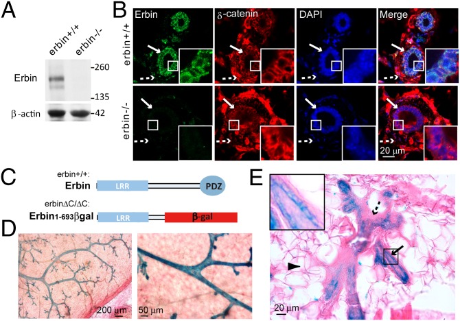
Fig. 1.
Erbin is expressed in luminal epithelial cells of mammary glands. (A) Expression of Erbin in mammary tissues. Mammary tissues of 3-mo-old mice were homogenized and blotted with affinity-purified anti-Erbin antibody. β-Actin served as loading control. (B) Localization of Erbin in luminal epithelial cells. Sections of mammary ducts were immunostained using anti-Erbin antibody. Arrows, luminal epithelial cells; dashed arrows, myoepithelial cells. (C) Schematic diagram of domain structures of wild-type Erbin and Erbin1–693βgal. Note that β-gal is expressed as a protein fused to truncated Erbin. (D) β-Gal activity was detected in mammary epithelial ducts in erbin+/ΔC mice. Mammary fat pads of erbin+/ΔC mice were stained whole mount for β-gal in situ activity. Shown are representative images at two magnifications. (E) β-Gal activity was confined to epithelial cells of the mammary gland from erbin+/ΔC mice. Arrowhead, adipocyte; Arrow, luminal epithelial cells; dashed arrow, myoepithelial cells. Areas in the small squares are enlarged in the Insets.

