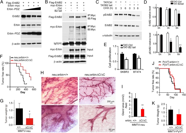
Fig. 6.
Requirement of the Erbin–ErbB2 interaction for ErbB2 stabilization and ErbB2-dependent tumorigenesis. (A) Decreased ErbB2 expression by the Erbin-PDZ domain. HEK293 cells were transfected with Flag-ErbB2 together with increasing doses of Myc-Erbin-PDZ. Cell lysates were blotted with antibodies against ErbB2 and Myc. (B) Disruption of the Erbin–ErbB2 interaction by B2tail. Lysates of transfected cells expressing Flag-ErbB2 or Myc-Erbin were incubated with control peptide (Ctrl) or B2tail. The Erbin complex was precipitated using anti-Myc antibody and probed for ErbB2. (C) Treatment with TAT-B2tail decreases ErbB2 stability and pErbB2 in human breast cancer cells. SKBR3 cells were incubated with 20 μM TAT-Ctrl or TAT-B2tail for 1 h before analysis of ErbB2 stability as in Fig. 5C. (D) Quantitative analysis of data in C (n = 3; *P < 0.05; **P < 0.01). (E) TAT-B2tail treatment inhibits proliferation of SKBR3 and BT474 cells. Cells were treated with 20 μM TAT-Ctrl or TAT-B2tail peptide for 24 h and analyzed by MTS assay (n = 3; *P < 0.05). (F) PDZ deletion inhibited tumorigenesis in MMTV-neu mice. neu;erbinΔC/ΔC and neu;erbin+/+ virgin littermates or sisters (n = 7 and 8, respectively) were examined weekly for tumors. Kaplan–Meier survival plot. Log-rank (Mantel–Cox) test, P < 0.0001. (G) Reduced tumor weight in neu;erbin ΔC/ΔC, compared with neu;erbin+/+ littermates. Tumors were isolated 5 wk after first detection of tumors (n = 7 and 8, respectively; *P < 0.05). (H) Reduced hyperplasia in nontumor mammary glands of neu;erbinΔC/ΔC mice. Shown is whole-mount hematoxylin staining of tumor-free mammary glands in neu;erbin+/+ and neu;erbinΔC/ΔC littermates during diestrus phase at the same age. (I) Quantitative analysis of data in H (n = 3; **P < 0.01). (J) PDZ deletion had no effect on tumorigenesis in MMTV-PyVT mice. PyVT;erbin+/+ (n = 23) and PyVT;erbinΔC/ΔC (n = 15) virgin littermates and sisters were examined for tumors. Kaplan–Meier survival plot. Log-rank (Mantel–Cox) test, P > 0.05. (K) PDZ deletion had no effect on breast tumor growth in MMTV-PyVT mice. Tumors were isolated 3 wk after detection (n = 8; P > 0.05).

