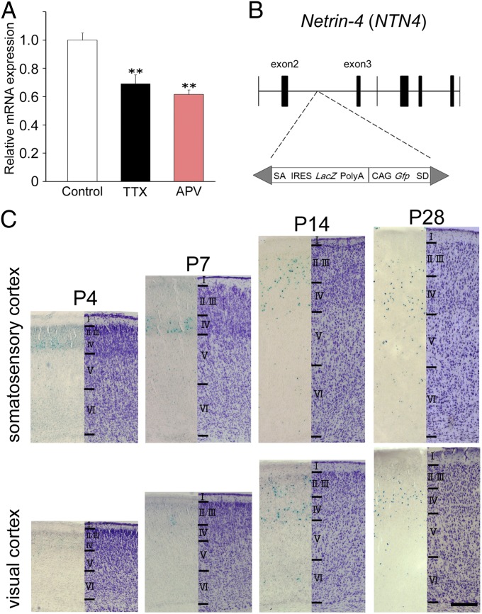
Fig. 1.
Expression of ntn4 in the cortex during development. (A) Ntn4 expression was examined by quantitative PCR in organotypic cocultures of the thalamus and cortex in normal culture medium (white) and in the presence of TTX (black) or APV (red). Histograms represent relative mRNA expression of ntn4 in cortical slices. Ntn4 expression was reduced by TTX as well as by APV application. Error bars show SEMs. **P < 0.01, Dunnett test. (B) Mutagenesis by insertion of the LacZ-containing cassette in ntn4 gene locus. SA, splice acceptor; IRES, internal ribosome entry site; SD, splice donor. (C) β-gal (Left) and Nissl staining (Right) in the somatosensory and visual cortex at P4, P7, P14, and P28. (Scale bar: C, 0.2 mm.)

