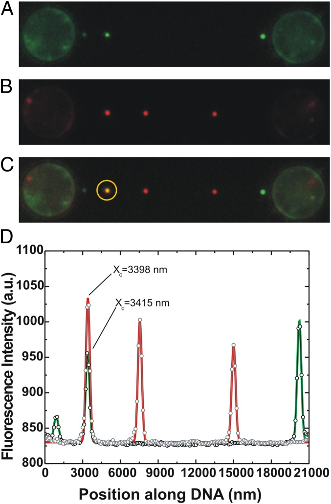
Fig. 2.
sm-FRAP allows detection of RAD51 growth on ssDNA. Conditions: RAD51 concentration, 12.5 nM; duration of an incubation period, 77 s. (A) Fluorescence image showing three individual fluorescent RAD51 nuclei on ssDNA. Subsequent continuous laser illumination resulted in complete photobleaching of the nuclei. (B) Fluorescence image of the same ssDNA–RAD51 complex after an additional incubation period. Fluorescent image shows the appearance of three distinct fluorescent patches. (C) Superposition of A and B allows the distinguishing of new nucleation events from RAD51 growth. In the yellow circle, we show that two of the fluorescent patches obtained from consecutive incubations colocalize exactly. (D) Line profile and Gaussian fitting of A and B confirm the colocalization of the two patches within 20 nm (fitted locations indicated by Xc). This confirms the direct separate detection of RAD51 nucleation and growth on ssDNA.

