Fig. 3.
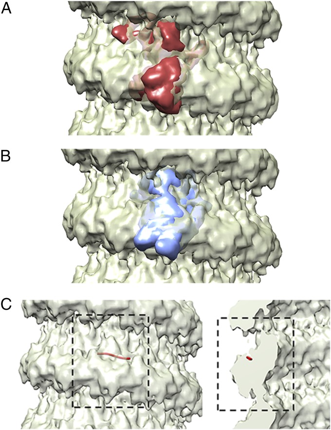
Comparison of MuV N to other NSV Ns. Comparisons of the MuV N to other NSV Ns was carried out by fitting simulated density maps from the crystal structures of VSV N-RNA complex (A; PDB ID code 2GIC) and RSV N-RNA complex (B; PDB ID code 2WJ8) into the MuV NC structure. The crystal structures are shown as simulated density maps (30, 45). The MuV N appears to share the typical structure as other NSV Ns, consisting of two core domains; NTD (Upper) and CTD (Lower). The density map of the MuV NC is presented at a sigma level of 2.5 (30) (C) The approximate location of the encapsidated RNA was determined by fitting the structure of a RSV N-RNA monomer (red string) into the MuV NC density. The viral RNA appears to be sequestered between NTD and CTD.

