Fig. 1.
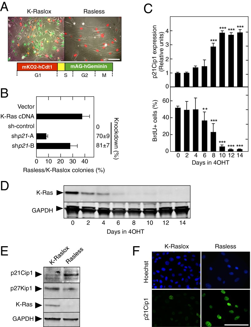
p21Cip1 is an essential mediator of cell cycle arrest in the absence of Ras signaling. (A) Cell cycle distribution of K-Raslox (Left) and Rasless (Right) MEFs stably expressing the FUCCI cell cycle indicators monomeric version of Kusabira Orange 2 (mKO2)/human chromatin licensing and DNA replication factor 1 (hCdt1) (red) and monomeric version of Azami Green (mAG)/human geminin (hGeminin) (green). (Lower) Schematic outline of the FUCCI cell cycle indicator system is shown. (B) Colony formation of Rasless and K-Raslox MEFs transfected with the indicated cDNA or shRNAs and expressed as the ratio between the number of colonies observed in cells lacking Ras proteins (Rasless) and those cells expressing K-Ras (K-Raslox). The knockdown efficiency of the shRNAs is indicated on the right side of the graph. Data are represented as mean ± SD. (C, Upper) qRT-PCR showing relative expression levels of p21Cip1 mRNA in K-Raslox MEFs treated for the indicated time with 4OHT. β-Actin expression levels were used for normalization. (C, Lower) Percentages of BrdU+ cells in K-Raslox MEFs treated for the indicated time with 4OHT. Data are represented as mean ± SD. **P < 0.01; ***P < 0.001 (unpaired Student t test). (D) Western blot analysis of K-Ras expression in K-Raslox MEFs treated for the indicated time with 4OHT using anti–Pan-Ras antibodies. GAPDH expression served as a loading control. (E) Western blot analysis of p21Cip1, p27Kip1, and K-Ras expression in K-Raslox MEFs either left untreated (K-Raslox) or treated with 4OHT (Rasless) for 2 wk. GAPDH expression served as a loading control. (F) Immunofluorescence staining of p21Cip1 expression in K-Raslox and Rasless cells. Cells were counterstained with Hoechst 33342 to visualize nuclei. (Scale bars, 100 μm.)

