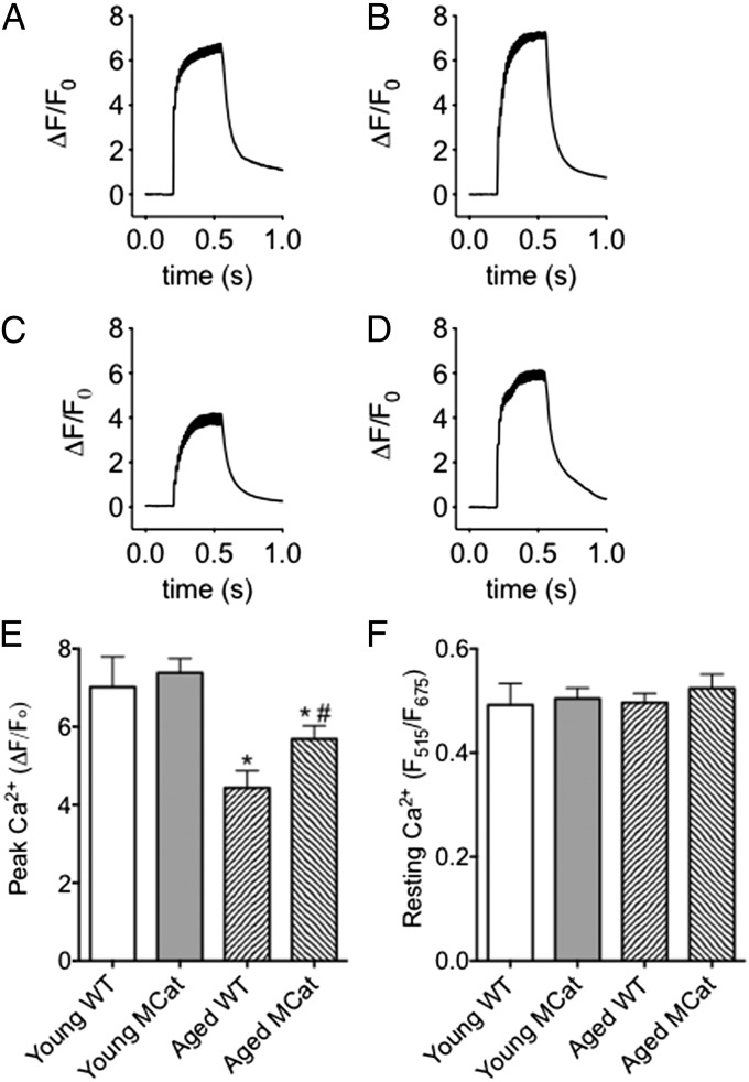
Fig. 3.
Improved tetanic Ca2+ in skeletal muscle from aged MCat mice. (A–D) Representative traces of normalized Fluo-4 fluorescence in FDB muscle fibers during a 70 Hz tetanic stimulation in young WT (A), young MCat (B), aged WT (C), and aged MCat (D). (E) Peak Ca2+ responses in FDB fibers stimulated at 70 Hz (fibers taken from the same animals as in A–D, n = 15–21 cells from at least three mice in each group). (F) Resting cytosolic Ca2+ (measured ratiometrically). Data are mean ± SEM (*P < 0.05 vs. young WT; #P < 0.05 vs. aged WT, ANOVA).

