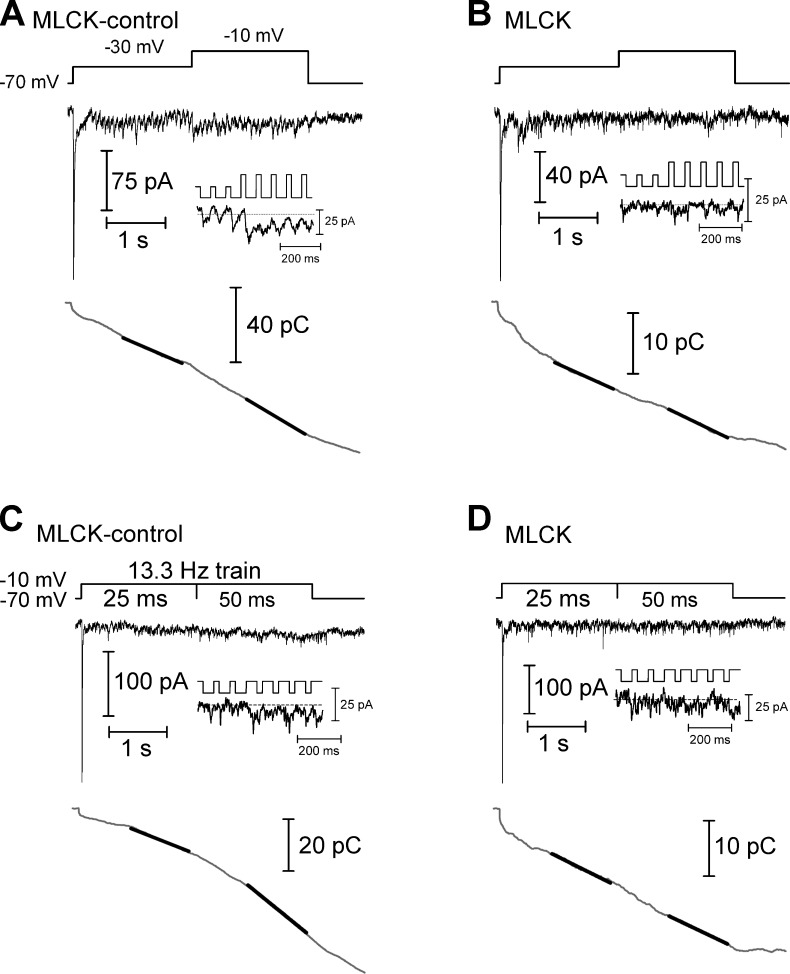
Figure 4.
Ca2+-dependent acceleration of replenishment measured using trains of depolarizing pulses. (A) In a paired recording of a cone and HC in which the cone was dialyzed with the MLCK-control peptide (20 µM), the cone was stimulated with a train of depolarizing pulses to −30 mV (25-ms duration, 13.3 Hz), evoking an EPSC in the HC (top). After 2 s, the step amplitude was increased to −10 mV to fully activate the Ca2+ current, accelerating replenishment. The inset shows the EPSC at the transition from steps to −30 mV to steps to −10 mV. The trace at the bottom shows the cumulative charge transfer obtained by integrating the EPSC waveform. The slope of a line (black line) fit to the final 1 s of the cumulative charge transfer of the EPSC in each experimental condition provides a measure of the rate of replenishment. (B) In a similar experiment, the cone was dialyzed with the MLCK peptide (20 µM). This inhibited the Ca2+-dependent acceleration of replenishment when the step amplitude was increased. (C) In a cone-HC paired recording in which the cone was dialyzed with the MLCK-control peptide, replenishment was accelerated when the depolarizing step (−70 to −10 mV) was lengthened from 25 to 50 ms. (D) This acceleration was inhibited when a cone was instead dialyzed with the MLCK peptide.

