Figure 1.
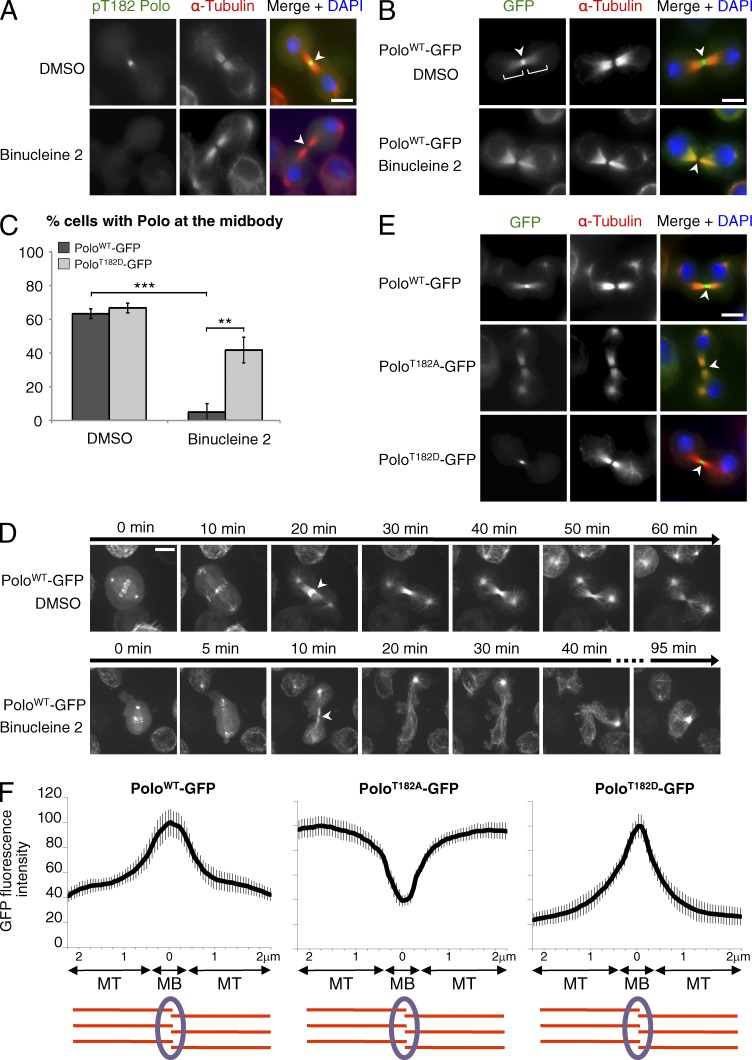
Phosphorylation of Polo by Aurora B regulates its localization in cytokinesis. (A) The localization of pT182-Polo at the midbody depends on Aurora B. Immunofluorescence in D-Mel2 cells is shown. Inhibition of Aurora B with Binucleine 2 reduced the pT182-Polo signal at the midbody (arrowheads). (B) The localization of Polo-GFP at the midbody depends on Aurora B. Binucleine 2 reduced the Polo-GFP signal at the midbody (arrowheads), but not the microtubule-associated pool of Polo-GFP (brackets). (C) Quantification of the number of cells showing clear localization of Polo-GFP or PoloT182D-GFP at the midbody after treatment with Binucleine 2 or DMSO (n = 20, repeated three times). Error bars indicate SD (**, P < 0.01; ***, P < 0.001; Student’s t test). (D) Imaging of Polo-GFP–expressing cells. Binucleine 2 or DMSO was added at anaphase onset (T0). Images were taken every 1 min. Arrowheads, midbody. (E) Mutation of Thr182 affects the localization of Polo in cytokinesis. Immunofluorescence in cells expressing PoloWT-GFP, PoloT182A-GFP, and PoloT182D-GFP is shown. (F) Line scans of the GFP fluorescence intensity along the intercellular bridge (n = 10, MT, microtubules; MB, midbody). Error bars indicate SD. Bars, 5 µm.

