Figure 1.

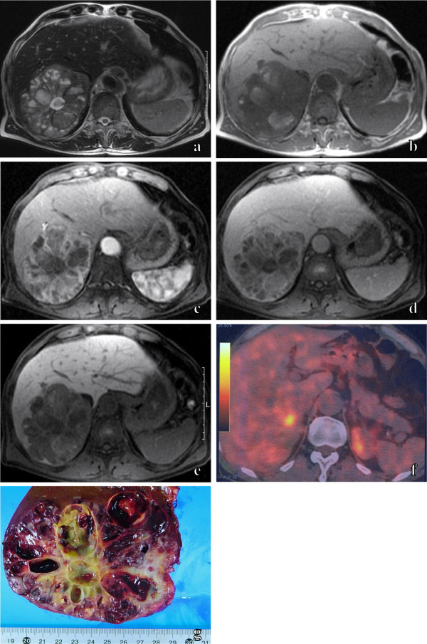
Primary hepatic carcinoid tumor in 70-year-old male. An axial T2-weighted MR image (a) shows the hepatic mass, which included multiple cystic areas with shading. An axial T1-weighted MR image (b) shows some hyperintense cystic areas in the hepatic mass. Axial fat-suppressed dynamic MR images with Gd-EOB-DTPA show the prolonged enhancement of solid areas and the non-enhancing multiple cystic areas of the hepatic mass from the early phase (c) to the late phase (d). An axial fat-suppressed MR image with Gd-EOB-DTPA in the hepatobiliary phase (e) shows the hypointense hepatic mass, the extent of which is clearly delineated by increasing the signal intensity of the normal liver parenchyma. 18 F-FDG PET/CT (f) shows no abnormal uptake of FDG in most of the hepatic mass, and increased uptake of FDG in the fraction of the hepatic mass. The cut surface of the resected specimen (g) shows a hepatic mass with multiple hemorrhagic cystic areas.
