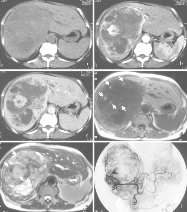
Figure 2.

Primary hepatic carcinoid tumor in 74-year-old female. Unenhanced CT (a) shows a heterogeneous hypodense mass mainly in the right lobe of the liver. Dynamic CT shows prolonged, enhancing, irregular solid areas and non-enhancing areas of the hepatic mass from the early phase (b) to the late phase (c). An axial T1-weighted MR image (d) shows the mainly hypointense mass, including scattered hyperintense cystic areas (arrows). An axial T2-weighted MR image (e) shows the mainly hyperintense mass, including cystic areas with shading (arrows). A celiac arteriogram in the early phase (f) shows an irregular tumor stain, which is supplied by the stretching right and middle hepatic arteries.
